Determining the role of protein quality control in neurodegeneration
Each cell is equipped with a protein quality control machinery, which includes molecular chaperones and cellular degradation pathways. This machinery maintains protein homeostasis (proteostasis), and prevents protein misfolding and aggregation. Protein quality control declines with age, leading to accumulation of damaged proteins and promoting the onset of neurodegeneration. However, how exactly proteostasis impairments contribute to the development of different neurodegenerative diseases remains elusive. To monitor neuronal proteostasis in vivo, our laboratory has generated a reporter mouse that expresses EGFP-fused firefly luciferase (Fluc-EGFP) in the brain (Blumenstock et al., 2021; Dudanova, 2022). Fluc is a conformationally unstable protein that requires chaperones for proper folding, and reacts to proteostasis disturbances by formation of intracellular Fluc-EGFP foci and by reduced luciferase activity (Fig. 1). With the help of these mice, we have found a marked impairment of proteostasis in the rTg4510 tauopathy mouse model, but not in the R6/2 Huntington’s disease model (Fig. 2), and showed that proteostasis impairments depend both on the identity of the aggregating protein as well as on its subcellular localization.
We are currently investigating proteostasis in other diseases and in different neuronal cell types. Moreover, the Fluc-EGFP mice enable us to observe the dynamics of proteostasis and protein aggregation during disease progression using chronic in vivo two-photon microscopy. These investigations will be important for predicting at which stage of disease the protein quality control system can be therapeutically targeted with the greatest success.
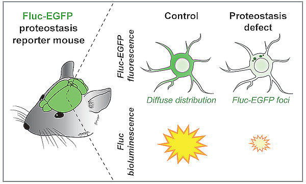
Figure 1.
Scheme of the proteostasis reporter mouse. Impairment of neuronal proteostasis can be detected by the appearance of Fluc-EGFP foci, and by reduced luciferase activity.

Figure 2.
Fluc-EGFP reporter reveals proteostasis impairment in tauopathy mice, but not in Huntington’s disease mice. Images show cortical neurons. Fluc-EGFP is in green, Tau and mutant Huntingtin (mHTT) aggregates in magenta, nuclei are labeled in blue. Arrowheads point to Fluc-EGFP foci. Adapted from Blumenstock et al., 2021.